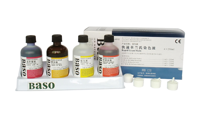
Rapid Gram Stain

Intended use:
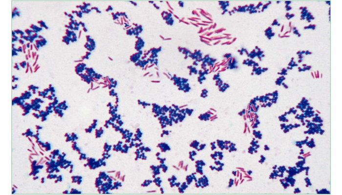
A rapid staining procedure to classify bacteria based on their cell wall characteristic. According to the stain results, bacteria can be divided into two different groups - Gram positive (purple) and Gram negative (red).
Principle:
Gram stain was devised empirically by Christian Gram, a Danish physician, in 1884. When bacteria are stained with certain basic dyes (such as the crystal violet in Rapid Gram Stain), those cells with high peptidoglycan contents (Gram-positive) in their cell wall will resist decolorization with organic solvents such as ethanol or acetone, while the Gram-negative species (with a thin layer of peptidoglycan in the cell wall) can be easily decolorized.
Methods:
1. Stain the sample slide with Crystal Violet for 10 seconds. Rinse with water gently, and then remove the
excess water.
2. Apply Iodine Solution for 10 seconds. Rinse with water gently, and then remove the excess water.
3. Add Decolorizer for 10~20 seconds. Rinse with water gently, and then remove the excess water.
4. Counterstain with Safranin Solution for 10 seconds. Rinse with water gently.
5. Blot up sample slide with bibulous paper (or allow to air dry), and then examine the slide under a microscope.
Specifications:
|
Contents |
4vialsx20ml/kit |
4Btlsx100ml/kit |
4Btlsx250ml/kit |
4x(6Btlsx500ml)/kit |
Components |
| Crystal Violet |
1x20ml |
1x100ml |
1x250ml |
6x500ml |
Crystal Violet, Ethanol |
| Iodine Solution |
1x20ml |
1x100ml |
1x250ml |
6x500ml |
Iodine, Potassium iodide |
| Decolorizer |
1x20ml |
1x100ml |
1x250ml |
6x500ml |
Acetone, Ethanol |
| Safranin Solution |
1x20ml |
1x100ml |
1x250ml |
6x500ml |
Safranin, Ethanol |
Precaution:
1. The length of stain will depend on the specimen type and smear thickness, etc. For female leucorrhea
smear, staining time should be slightly longer (more than 10 seconds) for better results.
2. Cap reagent bottle immediately after each use to prevent reagent evaporation.
3. Do not use any reagent beyond the stated expiration date. For kit storage, avoid exposure to extreme high
or low temperature and sunlight.
Expected Results:
Gram-positive organisms appear purple.
Gram-negative organisms appear red.


