
Leukocyte Peroxidase (POX) Stain
(Myeloperoxidase)
Intended Use:
This kit is for staining bone marrow cell and bloodcell smear.
Principle:
Peroxidases in cells dissolve oxides to generate oxygen. Oxygen shall react with potassium iodide to produce iodine. Iodine works with Wright Giemsa stain and forms colored granules in cytoplasm.
Specifications:
|
Contents |
5Tests/Kit |
20Tests/Kit |
100Tests/Kit |
Components |
|
Solution A |
1vial×2.5ml |
1vial×10ml |
1vial×50ml |
Eosin |
|
Solution B |
1vial×5ml |
1vial×20ml |
1vial×100ml |
Azure II |
|
KI PBS |
1vial×5ml |
1vial×20ml |
1vial×100ml |
potassium iodide |
|
Wright-giemsa |
1vial×1.5ml |
1vial×5.5ml |
1vial×25ml |
Wright-giemsa |
Methods:
Working solution Preparation (for 1 test only):
Apparatus required:disposable tube, micropipette, disposable tips, dropper;
Instruction:Shake the tube while adding 1.0ml solution C to 250µl solution D. Use the solution within 2 hours.
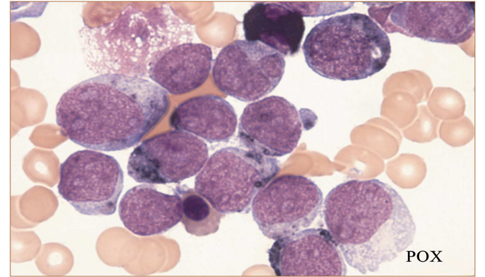
Expected Results:
In positive reaction, red-brown to dark blue granules shall be found in cytoplasm. Red-brown granules are with weakly positive reaction. Dark purple to dark blue granules spread over cytoplasm and even fully covernuclei in strong positive reaction. Blue cytoplasm means negative, and no positive granules are found, while the nuclei shall be evenly stained as red-purple. Eosinophils can be quickly stained in vivid color,and its positive reaction is dark blue. Some cellular granules shall spread diffusely to the outside of cell. Acicula shall appear in the outer layer of cells.